Syn.Flex.GCaMP6f.WPRE.SV40 (AAV9) in nucleus accumbens shell (NAcSh) brain slices from D1-Cre and A2A-Cre mice
Addgene #100833-AAV9
- Data Submitted By
- Sonia Ortega-Tinoco
- Lab Name
- Salvador Hernández-López, Departamento de Fisiología, Facultad de Medicina, Universidad Nacional Autónoma de México (UNAM)
- Submission Date
- September 08, 2025
- Publication Date
- October 17, 2025 (modified October 20, 2025)
- DOI
- https://doi.org/10.57733/addgene.a8yf1k
- Funding
- CONAHCyT 1102522
- Abstract
- Pituitary adenylate cyclase-activating polypeptide (PACAP) modulation of medium spiny neuron (MSN) activity was investigated through whole-cell recording and calcium imaging of nucleus accumbens shell (NAcSh) brain slices from D1-Cre and A2A-Cre mice. D1 and D2 medium spiny neurons expressing GCaMP6f were labeled via transduction with 100833-AAV9: pAAV.Syn.Flex.GCaMP6f.WPRE.SV40.
- Citation
-
Syn.Flex.GCaMP6f.WPRE.SV40 (AAV9) in nucleus accumbens shell (NAcSh) brain slices from D1-Cre and A2A-Cre mice. Ortega-Tinoco S 2025. Addgene Report, https://doi.org/10.57733/addgene.a8yf1k
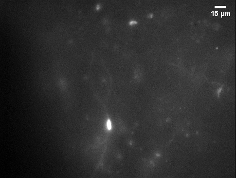
Image attribution: Sonia Ortega-Tinoco
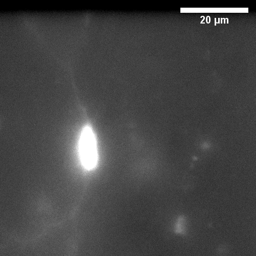
Image attribution: Sonia Ortega-Tinoco
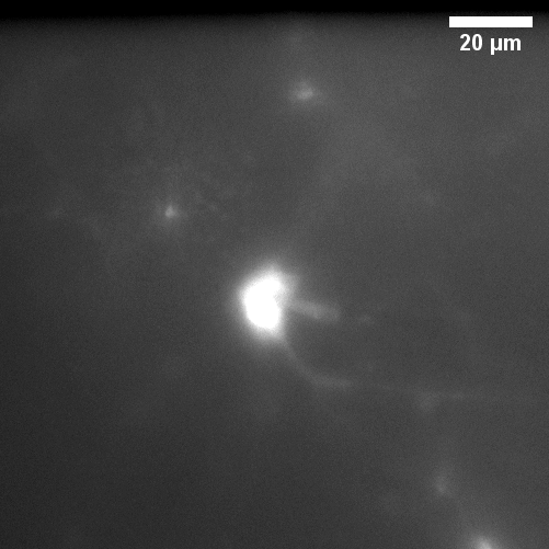
Image attribution: Sonia Ortega-Tinoco
This report is made available under the Creative Commons Attribution 4.0 International License.
- Vector Used
-
pAAV.Syn.Flex.GCaMP6f.WPRE.SV40
Addgene #100833-AAV9
Virus & Injection
Virus
- Virus Name
- pAAV.Syn.Flex.GCaMP6f.WPRE.SV40
- Serotype
- AAV9
- Source
- Addgene
- Catalog Number
- 100833-AAV9
- Promoter
- Synapsin
- Cargo Type
- Biosensor
- Injection Titer / Dose
- Bilateral injections of 0.6 μL of 100833-AAV9: pAAV.Syn.Flex.GCaMP6f.WPRE.SV40 were administered into the nucleus accumbens shell (NAcSh) using a 30G dental needle attached to a microinjection system, at a rate of 0.1 μL/min.
- Injection Volume
- 0.6 μL
- Injection Rate
- 0.1 μL/min
- Injector Material
- 30G dental needle
- Injection Site / Route
- Nucleus accumbens shell (NAcSh) at stereotaxic coordinates relative to bregma: AP +1.1 mm, ML ±1.1 mm, DV -5.2 mm.
Other Details
- Species
- Mouse
- Strain or Cell Line
- C57BL/6J, D1-Cre mice (Drd1a-Cre), and D2-Cre mice (Adora2a-Cre)
- Source
- D1-Cre mice: Jax stock #030329; D2-Cre mice: MMRRC_031168-UCD
- Age at Injection
- 30 days
- Sex
- Male
- Time After Injection
- 18–21 days
- Detection Method
- Whole-field fluorescence
- Assay & Results
- The image field was visualized under a microscope and the field size was 280 ×260 μm size. The tissue was stimulated with light pulses (488 nm wavelength, 15–25 ms exposure time) using a LED lamp (Lambda HpX L5, Sutter Instruments) connected to the microscope via fiber optics. Image sequences were acquired at a frequency of 4 frames/s (250 ms/frame) by using a digital camera (Cool SNAP MYO, Photometrics)
- Tips & Comments
- Please see the original publication for more details: Ortega-Tinoco, S., Padilla-Orozco, M., Hernández-Vázquez, F., Garduño, J., Mondragón-García, A., Ramírez-Sánchez, E., Bargas, J., & Hernández-López, S. (2025). PACAP induces increased excitability in D1- and D2-expressing nucleus accumbens medium spiny neurons. Brain research bulletin, 224, 111323. https://doi.org/10.1016/j.brainresbull.2025.111323