
Flow Cytometry for Anti-CELSR1 [IPI-mCELSR1.63]
Addgene #262534
- Data Submitted By
- Zachary Anderson, Haiying Li, Travis Riedel, Haisun Zhu and Deborah Moshinsky
- Lab Name
- Institute for Protein Innovation (IPI)
- Submission Date
- October 20, 2026
- Publication Date
- October 21, 2026 (modified November 12, 2026)
- Abstract
- Experimental results for Anti-CELSR1 [IPI-mCELSR1.63] (Addgene #262534) in a flow cytometry assay with ExpiCHO cells expressing mouse CELSR1.
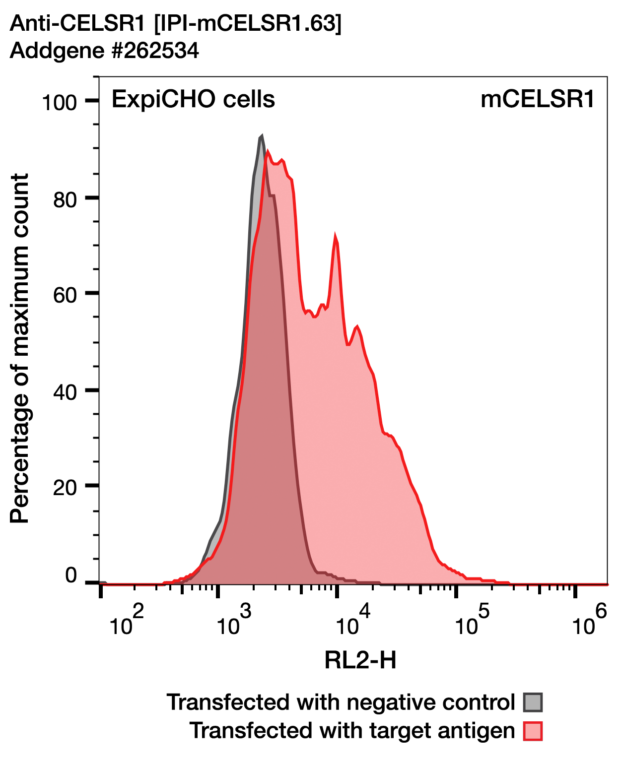
Image attribution: Institute for Protein Innovation (IPI)
This report is made available under the Creative Commons Attribution 4.0 International License.
- Antibodies Used
-
Anti-CELSR1 [IPI-mCELSR1.63]
Addgene #262534 -
Alexa Fluor 647 F(ab')2 goat anti-rabbit IgG Fc fragment
Jackson ImmunoResearch #111-606-046
Materials & Methods
Sample
- Target Species
- Mouse
- Cell / Tissue Type
- ExpiCHO cells
- Additional Information
- ExpiCHO cells were transiently transfected with mouse CELSR1 or B7H3 (negative control).
Buffer
- Name
- PBS
Primary Antibody
- Name
- Anti-CELSR1 [IPI-mCELSR1.63]
- Source
- Addgene
- Catalog Number
- 262534
- Host Species
- Rabbit
- Target Antigen
- CELSR1
- Concentration
- 15 µg/mL
- Time
- 30 min
- Temperature
- On ice
Secondary Antibody
- Name
- Alexa Fluor 647 F(ab')2 goat anti-rabbit IgG Fc fragment
- Source
- Jackson ImmunoResearch
- Catalog Number
- 111-606-046
- Concentration
- 2 µg/mL
- Time
- 30 min
- Temperature
- On ice
Additional Information
- Additional Information
EC50: A fourteen point titration of antibody concentrations, ranging from 660 nM (0.1 mg/mL) to 4.42 pM with a 1:2.5 dilution factor, against mouse CELSR1 showed reactivity towards mouse CELSR1 with an observed EC50 value of 0.77 nM.
Protocol: ExpiCHO cells were grown per manufacturer’s instruction. Plasmids expressing mouse CELSR1 were transiently transfected into cells together with a plasmid expressing blue fluorescent protein (BFP). Control cells were transfected with plasmid expressing full-length human B7H3 together with the BFP plasmid. Two days after transfection, cells at a density of 3 x 10⁶ cells/mL were labeled with primary antibodies.
- Primary antibody: 15 µg/mL in 1 mL Dulbecco's phosphate-buffered saline (DPBS), 0.5% BSA, with 0.9 mM Ca2+, 0.5 mM Mg2+, kept at 4 °C. 50 µL of antibody added to cells and incubated on ice for 30 min.
- Washing conditions: Cells pelleted and washed twice in 0.5% DPBS, 0.5% BSA, with 0.9 mM Ca2+, 0.5 mM Mg2+.
- Secondary antibody: 2 µg/mL Alexa Fluor 647 F(ab')2 goat anti-rabbit IgG Fc fragment (Jackson ImmunoResearch, 111-606-046) in 0.5% DPBS, 0.5% BSA, with 0.9 mM Ca2+, 0.5 mM Mg2+, 30 min, on ice.
- Detection: Labeled cells were analyzed with an Intellicyt iQue Screener Plus flow cytometer. Cells were first gated for positive BFP expression and then gated for the Alexa 647-conjugated secondary antibody.
- Controls
- Control cells were transfected with plasmid expressing full-length human B7H3 together with the BFP plasmid.
Results
- Result
- Pass (The antibody worked under these conditions)
- Describe Results
- Recognizes mouse CELSR1.