Anti-Neurexin-3-alpha [IPI-mNRXN3a.67] in immunohistochemistry (IHC) (Mouse)
Addgene #237694
- Data Submitted By
- Alexander Morano , Travis Riedel and Deborah Moshinsky
- Lab Name
- Institute for Protein Innovation (IPI)
- Submission Date
- May 29, 2026
- Publication Date
- June 10, 2026 (modified June 18, 2026)
- DOI
- https://doi.org/10.57733/addgene.3vufg5
- Abstract
- Experimental results for Anti-Neurexin-3-alpha [IPI-mNRXN3a.67] (Addgene #237694) in an immunohistochemistry (IHC) application on mouse hippocampus.
- Citation
-
Anti-Neurexin-3-alpha [IPI-mNRXN3a.67] in immunohistochemistry (IHC) (Mouse). Morano A, Riedel T, Moshinsky D 2026. Addgene Report, https://doi.org/10.57733/addgene.3vufg5
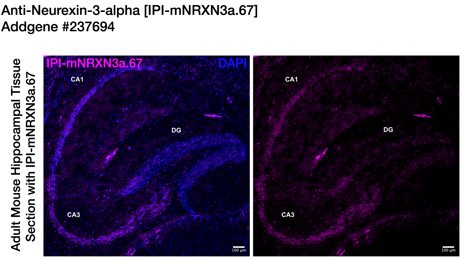
Image attribution: Institute for Protein Innovation (IPI)
This report is made available under the Creative Commons Attribution 4.0 International License.
- Antibodies Used
-
Anti-Neurexin-3-alpha [IPI-mNRXN3a.67]
Addgene #237694 -
Goat anti-rabbit alexa fluor 647
ThermoFisher #A21245
Materials & Methods
Sample
- Target Species
- Mouse
- Cell / Tissue Type
- Hippocampus
- Additional Information
- P35 C57BL/6J mouse brains
Buffer
- Name
- PBS
Fixative
- Fixative
- Paraformaldehyde, 4% in PBS
- Time
- 1.5 h post-fixation with PFA after perfusion and harvesting
- Temperature
- 4 ˚C
Permeabilization / Delipidation
- Agent
- PBS with 0.1% Triton-X 100
- Time
- 15 min (3x 5 min washes)
- Temperature
- Room Temperature
Blocking
- Agent
- Normal Goat Serum
- Concentration
- 5% in PBST
- Time
- 1 h
- Temperature
- Room Temperature
Primary Antibody
- Name
- Anti-Neurexin-3-alpha [IPI-mNRXN3a.67]
- Source
- Addgene
- Catalog Number
- 237694
- Host Species
- Rabbit
- Target Antigen
- NRXN3
- Concentration
- 10 µg/mL (1:100 dilution)
- Time
- 60 min
- Temperature
- Room Temperature
Secondary Antibody
- Name
- Goat anti-rabbit alexa fluor 647
- Source
- ThermoFisher
- Catalog Number
- A21245
- Concentration
- 1:500 dilution from a 2 mg/mL stock
- Time
- 90 min
- Temperature
- Room Temperature
Additional Information
- Additional Information
Immunohistochemistry Methods: Mouse brain tissue
Adult (3–5 week old) mouse brains were first purchased from Jackson Labs. Instructions were provided to Jackson labs for harvesting and fixing tissue. Anesthesia was performed with inhalant isoflurane, followed by transcardial perfusion with ~11 mL 1X PBS perfusate/mouse containing heparin (10,000 units heparin per 1L 1X PBS), then ~25 mL ice-cold 4% paraformaldehyde in 1X PBS. After dissecting the brain, it was stored in 1X PBS + 0.01% sodium azide solution (NaN3) at 4 °C. Upon arrival of tissue, adult brains were placed in a 5 mL conical tube containing ice-cold (4 °C) 10% sucrose solution and incubated at 4 °C overnight. The next day, 10% sucrose was replaced with ice-cold 30% sucrose and the brain was allowed to equilibrate completely at 4 °C, as evidenced by its sinking to the bottom of the tube, for 2–3 days.
Equilibrated tissue was then frozen in Optimum Cutting Temperature (OCT) on dry ice and 20–25 μm sections were cut on a Leica cryostat at the Harvard histology/sectioning core. Slides were thawed for 5 min at RT; sections were rehydrated 1x 5min in 1X PBS and then washed 3x 5 min in 1X PBST (0.1% Triton X-100 in 1X PBS) in a humidified chamber in a slide box. Slides were blocked for 1 h at RT in 5% NGS (Normal Goat Serum) or 5% NDS (Normal Donkey Serum) in 0.1% Triton-X-100 in PBS. Primary antibodies were then diluted in blocking solution and incubated overnight at 4 °C. Slides were then washed 4x 5min in 0.1% TritonX-100 and incubated with secondary antibodies diluted in blocking solution (5% NGS or NDS) at RT for 1.5–2 h, in the dark. Slides were washed 4x 5 min in 0.1% TritonX-100 again, then 1x 5min in 1X PBS. Slides were then mounted in Fluoromount mounting medium with DAPI, outlined with nail polish to secure the coverslip, and stored at 4 °C in the dark until ready to image.
Controls
Negative control: No primary control.
Results
- Result
- Pass (The antibody worked under these conditions)
- Describe Results
- This antibody shows strongest labeling in the CA1 and CA3 regions of the hippocampus, aligning with expression data detailed in the Allen Brain Atlas. Further validation in genetic knock-outs is recommended to confirm specificity of the signal.